Research Goals
the research goals of CAPS try to objectively access the subjective surgical assessment criteria of body contouring changing operations using 3-D patient assessment. Based on these objective findings of different innovative 3-D technologies a more objective evaluation of the surgical outcome is possible. The research group CAPS intends to apply established industrial simulation techniques in the field of Computer Aided Engineering (CAE) to specific medical questions and to develop clinical applications. Our challenge is to establish a quality assurance in the field of Plastic, Reconstructive and Aesthetic Surgery in terms of an evidence-based plastic surgery and to develop more transparent assessment criteria of competing surgical treatments.
3-D Measurement
Many years of experience in optimization, standardization and validation of different 3-D optical assessment technologies for the quantification of surgical induced shape and volume changes of various anatomical regions. Identification of clinical relevant applications and establishment of different, noninvasive, optical 3-D scanning technologies to clarify specific medical questions.
3-D Visualization
Development of methods for the generation of patient specific, virtual, anatomical 3-D models with the help of reconstruction and registration of various image generation modalities (e.g. computed tomography, magnetic resonance imaging, 3-D ultrasound imaging, optical 3-D scanning technologies, holographic capturing). Generation of mathematically precise 3-D models for computer aided surgical planning and simulation by using segmented radiological image data as well as rapid prototyping - / manufacturing technologies.
Development of Force-Feedback Applications (Haptic Modeling)
Standardization and validation of interactive 3-D morphing systems for intuitive haptic modeling of virtual 3-D models. Identification of clinical applications for computer aided surgical planning using force-feedback systems without taking into account the biomechanical behaviour of the human tissue.
Finite Element Simulation
Based on the 3-D reconstruction of virtual anatomical 3-D models from diffrent anatomical regions, the implementation of the biomechanical tissue parameters is performed in order to run numerical simulations of the soft tissue deformation by the use of the Finite Element Method (FEM). By using this method deformable FEM models can be generated for the simulation and valdiation of biomechanical interactions with respect to the realistic and specific tissue parameters. In this context the relevant physical tissue parameters are identified. These are highly relevant for the numerical simulation.
Biomechanics
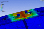
With the help of numerical simulation the validation of established biomechanical analysis methods is performed. Based on FEM (Finite Element Method) the biomechanical interaction of relevant medical questions can be simulated, visualized and quantified. One goal of the cooperation with the Department of Traumatology of the Klinikum Rechts der Isar is the comparison of competing concepts, optimization and improvement of various technologies in osteosynthesis.


